Solid state 31P and 1H micro-imaging in 2D and 3D with magic angle spinning on biological sample and biomaterials
- 1. CEMHTI-CNRS, Orléans, France
- 2. Leibniz-Institut für Polymerforschung, Dresden, Germany
MRI is a well established non-invasive technique allowing 3D imaging of soft tissues. However, in case of rigid tissues, it suffers from several limitations: the short transverse relaxation time (T2) prohibits the use of spin echo MRI sequences and the wide resonances decrease both the sensitivity and the resolution obtained with frequency encoding. To overcome these drawbacks, the Magic Angle Spinning (MAS) can be used to remove the anisotropic broadening and to increase the T2 dephasing time in rigid solids.
The potential of MAS-MRI to obtain 31P and 1H images of rigid biological tissues is demonstrated on a mouse tooth with a piece of the attached jaw bone. We have employed a very high magnetic field (17.6 T) to increase the sensitivity and thus the spatial resolution. The 31P and 1H images have been recorded at a MAS frequency of 10 kHz using spin echo MRI sequences. In the experiments, the phase encoding gradients were designed to follow the rotation of the rotor, allowing to obtain static images of the rotating sample.
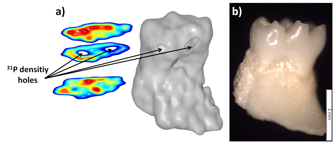
31P images of the mouse tooth and bone were recorded using cross-polarization to increase the signal and also to decrease the repetition time. The 31P 3D image exhibits spatial resolution of 220 µm in the phase dimensions and 280 µm in the read dimension. This allows observing two lacks of phosphorus density in the tooth which correspond to the pulp channels.

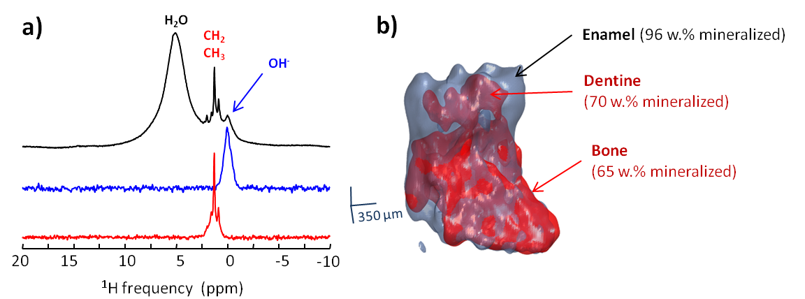
Two 1H images of the same sample have been recorded with the same resolution. Thanks to the magic angle spinning, the proton spectrum showed three different components (figure 2). 1H images of the hydroxyl hydroxyapatite resonance (in blue) and of the proteins aliphatic zone (in red) have been selectively recorded. The comparison of these two 1H images allows to discriminate different zones in the sample associated to variations of the mineralization degree. In particular, this contrast allows a clear distinction between the enamel and the dentine part of the tooth.

MAS-MRI can provide non-invasive 31P and 1H images of biological rigid tissues with a resolution up to 100 µm. This resolution is far below the one obtained in Micro Computed Tomography. However MAS-MRI provide a wide range of contrast possibility resulting from spectral selectivity or from some solid state NMR interactions such as the heteronuclear dipolar coupling expressed in the Cross Polarization sequence.
