Insights into fouling behavior of hollow fiber membrane modules
- RWTH Aachen University, Institut für Technische und Makromolekulare Chemie, Aachen, Germany
Filtrations play a tremendous role in humans' everyday life, for instance for treating drinking water/coffee/beer or driving cars. Although these are common tasks in most peoples' daily routine, the fouling behavior of filters and membranes is not yet fully understood. Typically the observed parameters are averaged over the whole membrane. These volume-averaged values are exploited to monitor membrane fouling, which leads to relatively vague conclusions. An example is the transmembrane pressure (TMP)[1], the pressure difference between the feed and the permeate solution, and steadily rising with the cake layer thickness.
In contrast MRI and MRI-velocimetry are able to give insights into the fouling behavior of membranes, as they can be slice-sensitive, do not require transparent systems and contrary to other techniques do not depend on cutting membranes open at different points in time in order to study the cake layer formation. There is plenty of literature available on fouling of reverse-osmosis (RO) membranes, flow-cells and hemodialyzers[2] [3] [4] but only very few on fouling of hollow fiber modules used for micro- & ultrafiltration of hard spheres[5] and especially microgels.

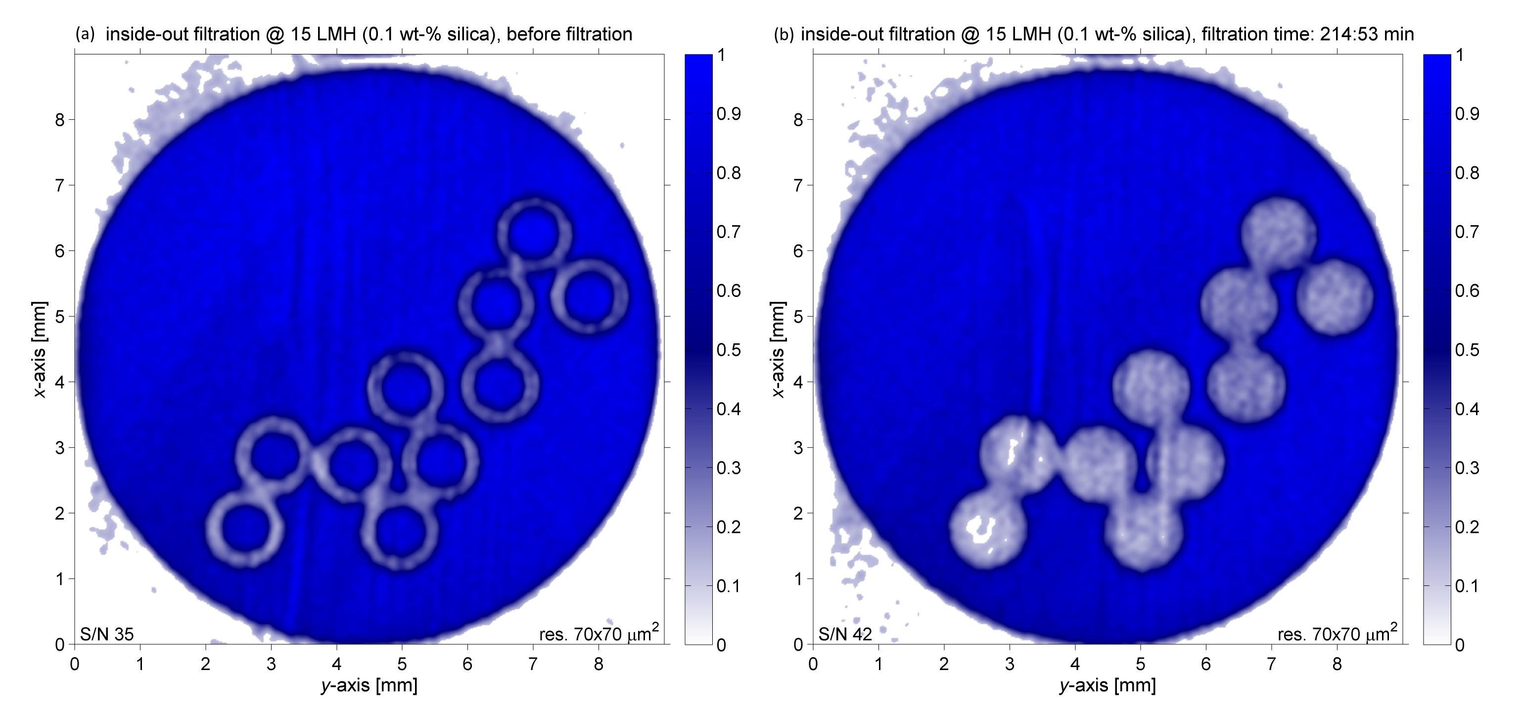
The study focuses on real-time MRI-velocimetry of silica ultrafiltration processes using hollow fiber membrane modules in dead-end inside-out as well as outside-in operation.Each module has an inner diameter (ID) of 9 mm and consists of ten membranes with an outer diameter (OD) of 1.2 mm and an ID of 0.7 mm. The membrane fouling was examined by using the sequence FLIESSEN (Flow Imaging Employing Single Shot Encoding),which is capable of measuring high-resolution (70·70 µm²/pixel) spin-density- as well as velocity-maps within 20 seconds, which can be considered real-time on the timescale of a filtration experiment. Figure 1 illustrates a membrane module before filtration (a) and towards the end of an inside-out filtration with Stöber silicates (mean hydrodynamic diameter 51.0 nm) (b). Initially the hollow fibers are solely filled with water, whereas the spin-density is significantly lowered once the membranes start clogging. The cake layer thickness at each point in time can be estimated by radial processing of each membrane, subsequent subtraction from the initial state and a count of pixels with decreased intensity.
- [1] Field RW, Wu D, Howell JA, Gupta BB, (1995), Critical flux concept for microfiltration fouling, Journal of Membrane Science, 100(3), 259-272
- [2] Graf von der Schulenburg DA, Vrouwenvelder JS, Creber SA, van Loosdrecht MCM, Johns ML, (2008), Nuclear magnetic resonance microscopy studies of membrane biofouling, Journal of Membrane Science, 323(1), 37-44
- [3] Laukemper-Ostendorf S, Lemke HD, Blümler P, Blümich B, (1998), NMR imaging of flow in hollow fiber hemodialyzers, Journal of Membrane Science, 138(2), 287-295
- [4] Hardy PA, Poh CK, Liao Z, Clark WR, Gao D, (2002), The use of magnetic resonance imaging to measure the local ultrafiltration rate in hemodialyzers, Journal of Membrane Science, 204(1-2), 195-205
- [5] Buetehorn S, Utiu L, Küppers M, Blümich B, Wintgens T, Wessling M, Melin T, (2011), NMR imaging of local cumulative permeate flux and local cake growth in submerged microfiltration processes, Journal of Membrane Science, 371(1-2), 52-64
