Hyperpolarisation and Biomedical MR II - L-034
Video-mode 3D imaging of rat lungs for critical-care medicine research
- 1. ABQMR, Albuquerque, United States
- 2. Brigham and Women's Hospital, Pulmonary and Critical Care, Boston, United States
- 3. Brigham and Women's Hospital, Laboratory of Mathematics in Imaging, Boston, United States
- 4. Brigham and Women's Hospital, Medicine, Boston, United States
- 5. Lovelace Respiratory Research Institute, Lung Cancer, Albuquerque, United States
- 6. Lovelace Respiratory Research Institute, Animal Care, Albuquerque, United States
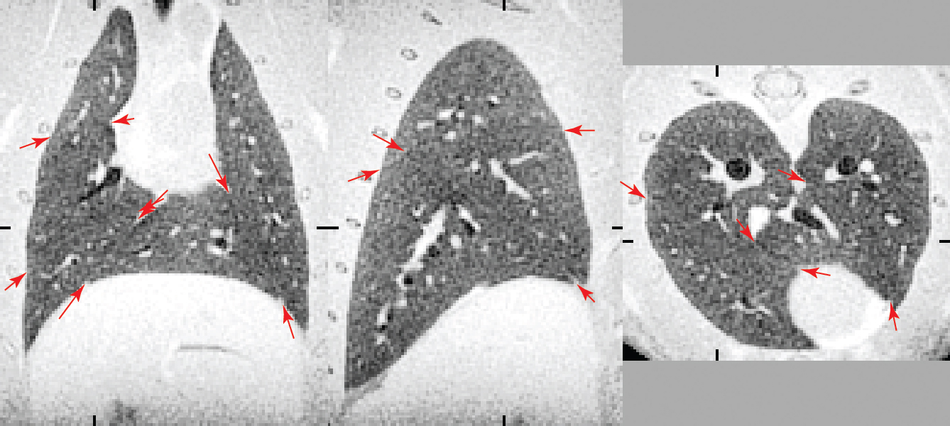
Lung parenchyma (the gas-exchanging, thin-wall tissue) is difficult to image because it is inhomogeneously broadened to about 8 ppm linewidth. It is typically black in MR images. FID-projection (now commonly called ZTE for "zero echo time") imaging can capture the short-lived signal. Visual criteria one can use for resolved lung parenchyma are: Lungs are lighter gray than airway lumens, which are black. They are lighter grey at exhaled volume than at inhaled volume. Fissures between lung lobes are visible when the image is of sufficient resolution. Fine airways and blood vessels are sharp and properly black and bright, respectively.

Coronal, sagittal, axial views of rat lung, 0.27 mm resolution. Red arrows show fissures between lung lobes. Airways are black; lung parenchyma is dark grey. Ticks show locations of the other views.
Success requires a high-dynamic-range receiver, low noise, rapid data transfer, large data sets, and precise synchronization with the ventilator. To extend the methods of [1] to overcome equipment limitations listed therein, we use a Tecmag Redstone spectrometer, with a Dell Precision T3600 computer (3.7 GHz clock). The 80 MHz NMR signal is directly digitized at 40 MHz to 16-bits to provide high dynamic range. Although Windows 7 is not a real-time operating system, the computer can keep up with 1 MHz data rate to store 10 GB data sets directly in 64 GB of RAM. This allows collecting 13 video frames of the respiratory cycle, with each image made from 120960 FIDs in a 25 minute scan time. A separate Macintosh Pro with 6 dual-core Xeon 3.5 GHz processors is used for image computation.
This system was developed primarily for the study of ventilator-induced lung injury (VILI) and acute respiratory distress syndrome (ARDS) which can be reproduced in rats to mimic the human ailments. A prominent feature is that lungs get infiltrated with edema and inflammatory cells. This puts more mechanical stress on the remaining lung tissue, which predisposes it to infiltration and a positive-feedback scenario can lead to asphyxiation. A challenge is to detect small regions of infiltration before they can be detected physiologically, which we are now able to do with MRI. Video-mode images at 0.4 mm resolution have been more useful than images at 0.27 mm resolution with more signal averaging. The resolution is sufficient for detecting small lesions and the change in grey scale between inhaled and exhaled lung volumes shows whether the region is still being ventilated. The visual dynamic gives a good impression of the animal's state and, with the parenchyma properly resolved, the grey scale of the images gives a quantitative measure of the degree of infiltration.
