Mapping Pressure Gradient Variation Using MRI and Microbubbles
- 1. Nottingham Trent University, School of Science & Technology, Nottingham, United Kingdom
- 2. University of Nottingham, Sir Peter Mansfield Magnetic Resonance Centre, Nottingham, United Kingdom
Introduction: Gas-filled microbubbles with lipid membranes have been used as a pressure sensitive probe in MRI [1] [2]. These bubbles undergo a radius change when an external pressure is applied, which consequently changes the external magnetic field perturbation and thus the T2 relaxation time [2]. The aim of this work is to use MRI to map a spatial pressure gradient using the microbubble contrast agent.
Methods: Monodisperse microbubbles with diameter in the range 2-3μm were prepared with a 1,2-dihexadecanoyl-sn-glycero-phosphocoline (DPPC) lipids shell and filled with C3F8 gas. The preparation was immobilized in a 1.5% Gellan gel suspending medium and loaded into a sample holder which was comprised of two compartments, separated by a 3mm thick sintered glass disc with 5-15μm pores to create a low permeability step between the two compartments. The pressure was applied to one compartment and further imaged using a Bruker Biospec 2.35T scanner with the RARE sequence.


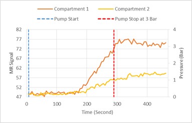
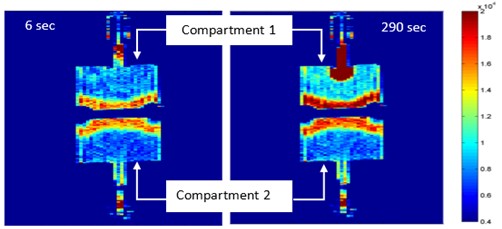
Results: Figure 1 shows the change in MR signal intensity due to the variation of pressure across the two compartments. The signal intensity in compartment 1 starts to increase 150 seconds after pump was turned on followed, 30 seconds later, by compartment 2. However, the signal in compartment 1 increases more rapidly which indicates a spatial pressure gradient between both compartments. As the pump stopped, the signal intensity in compartment 1 decreased whilst in compartment 2 it continued to rise, indicating the pressure is still equilibrating. Figure 2 shows two MR images of both compartments taken at different times and also shows the spatial pressure gradient.
Conclusion: A spatial pressure gradient on microbubbles is demonstrated by MR imaging in this preliminary study. We show that the pressure and its time course are different in both compartments. We will also show the time taken for the pressure to reach equilibrium for separating porous media with different permeabilities and match the MR signal intensity changes to pressure for a longer time of experiment. This work shows a great potential use of microbubbles contrast agents to measure intracardiac pressure changes and gradient pressure across a vascular stenosis using MRI.
- [1] Bencik M, Alrwaili A, Morris R, Fairhurst DJ, Mundell V, Cave G, McKendry J & Evans S, (2012), Quantitation of MRI Sensitivity to Quasi-Monodisperse Microbubble Contrast Agents for Spatially Resolved Manometry, Magnetic Resonance in Medicine, 1409-18
- [2] Alexander AL, McCreery TT, Barrette TR, Gmitro AF, Unger EC, (1996), Microbubbles as Novel Pressure-sensitive MR Contrast Agents, Magnetic Resonance in Medicine, 801-806
