New insights into membrane fouling by sodium alginate
- 1. Karlsruhe Institute of Technology, Institute of Mechanical Process Engineering and Mechanics, Karlsruhe, Germany
- 2. Karlsruhe Institute of Technology, Pro2 NMR, Institute f. Biological Interfaces 4, Institute of Mechanical Process Engineering and Mechanics, Karlsruhe, Germany
- 3. MANN+HUMMEL GmbH, Ludwigsburg, Germany
Fouling is one of the critical issues affecting the productivity, plant operation and maintenance costs of membrane bioreactors (MBR), one of the main causes being extracellular polymeric substances (EPS). Sodium alginate (SAL) often serves as a model compound for EPS. In the presence of divalent cations alginates form complexes.
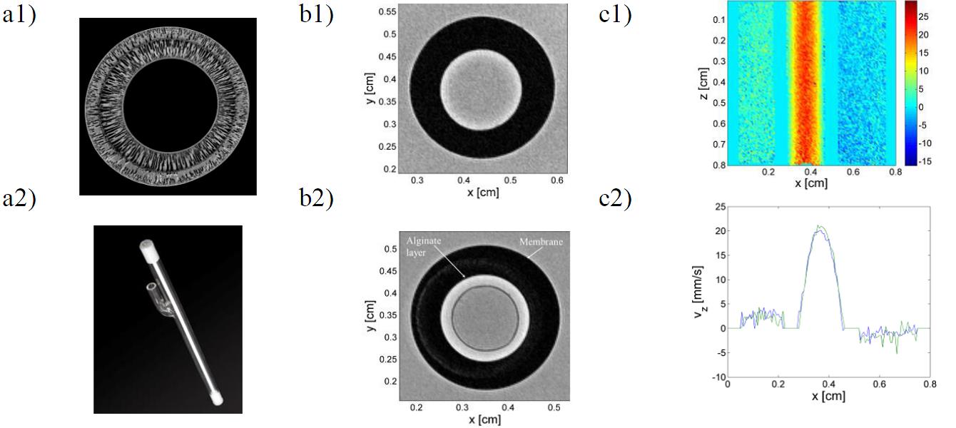
Operational parameters and membrane material have a large influence on the fouling behavior. Ceramic hollow fiber membranes (CHFM) are characterized by high chemical, thermal and mechanical stability as well as a high specific membrane filter surface. CHFM were investigated with respect to their fouling behavior, using SAL as model compound. The studied membranes have an inner diameter of 1.8 mm and consist of two layers: a support layer and a thin active layer of Al2O3, located at the inner side of the membrane. Results from filtration experiments suggest that the addition of calcium ions to the feed leads to a dense gel layer on the membrane surface. In order to investigate the resulting structures in the CHFM, we performed MRI measurements in addition to filtration experiments. To enhance contrast between alginate layer and surrounding water, we applied condensed clustered magnetic iron oxide nanocrystallites (MION) containing sodium alginate [1] [2] . After adding the contrast agent MION it is possible to identify an alginate gel-layer on the membrane surface (Fig. 1.b1). Long time experiments show additionally the diffusion potential of the SPION moieties in the filter.

The MRI measurements confirm the results obtained from dead-end filtration experiments.
To visualize the flow inside the membrane we performed FLOW PC measurements (Fig. 1.c) which reveal the mass transport in the filter module.
