Optimising multiple pulse arterial spin labelling for rodent brain imaging
- 1. University of Glasgow, Institute of Neuroscience and Psychology, Glasgow, United Kingdom
- 2. University of Glasgow, Institute of Cancer Sciences, Glasgow, United Kingdom
Arterial Spin Labelling techniques (ASL) measure tissue blood flow by manipulating the magnetization of arterial blood so as to distinguish it from that of static tissue. The ability to characterise non-invasively cerebral blood flow makes ASL a precious tool in the assessment of a wide range of pathologies, as stroke, brain tumour or epilepsy. The efficient spin labelling provided by a continuous ASL (CASL) is counter-balanced by the need for an extra-coil applying a continuous RF pulse at the carotid region and important magnetisation transfer effects at higher magnetic fields. In Pulsed ASL (PASL), continuous RF labelling is replaced by a pulsed RF labelling prior to the image acquisition. In the case of rodent imaging, such techniques are limited to low SNR, even when tagging is performed by whole-body volume coil and signal reception with a surface reception coil. A multiple inversion pulse sequence was proposed by Moffat et al [1] in order to address this limitation, which consists in tagging with a train of adiabatic HS inversion pulses located below the image plane. Their study showed improved SNR compared to standard, single pulse ASL.
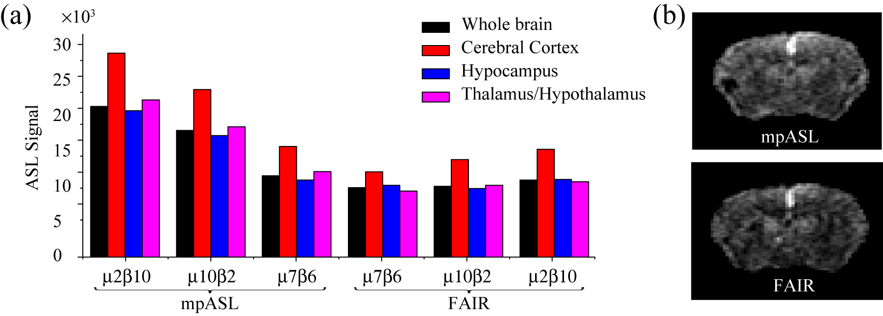
In this work, we propose a systematic approach in order to maximise the multiple pulse ASL (mPASL) signal. This involves the adjustment of several parameters going from the shape of the adiabatic RF pulses (Fig.1a) to the time frame of the sequence. mPASL optimisation was evaluated on mice and rat brain imaging. mPASL was compared with the commonly used Flow-sensitive Alternating Inversion Recovery technique (FAIR) (Fig.1b), that combines slice-selection and global inversion [2] . We show that the performance of each technique is depending on the animal to be scanned, with the animal length and weight playing important roles. The resulting signal is also shown to depend on the region of interest, with each technique exhibiting different signal distribution across brain regions. We discuss strategies for choosing the optimal sequence and parameters.

