Flow and Diffusion Magnetic Resonance I - L-050
Metrics of microscopic anisotropy: a comparison study
- 1. UCL, London, United Kingdom
- 2. Champalimaud Centre for the Unknown, Lisbon, Portugal
Introduction: Non-invasive estimation of cell size and shape is a key challenge in diffusion MRI. This work compares in simulation two recently introduced metrics of microscopic anisotropy: fractional eccentricity (FE), derived from double-diffusion-encoding (DDE) sequences with parallel and perpendicular gradients [1] (Method 1) and microscopic fractional anisotropy ( μ FA), derived from a combination of isotropic and directional gradients [2] (Method 2). We investigate two types of microscopically anisotropic substrates, which feature either restricted diffusion or Gaussian diffusion.

Diffusion sequences and substrates
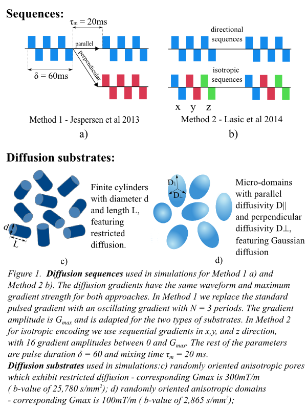
Methods: We construct diffusion sequences which are suitable for the acquisition techniques described in [1] and [2] . The sequences which are illustrated in Figure 1a) and 1b) have the same gradient waveform and maximum gradient strength, adapted for each substrate. We investigate two types of microscopically anisotropic diffusion substrates featuring either restricted diffusion or Gaussian diffusion, as illustrate in Figure 1c) and 1d). To compute FE, we derive the b-value and q-value for the sequences in Figure 1a) and use the equations from [1]. To compute μFA, we fit the cumulant expansion with the same mean diffusivity and different variances to the directional average and isotropic data. We find this to be more numerically stable than a Gamma distribution of diffusivities.

Microscopic anisotropy metric comparison
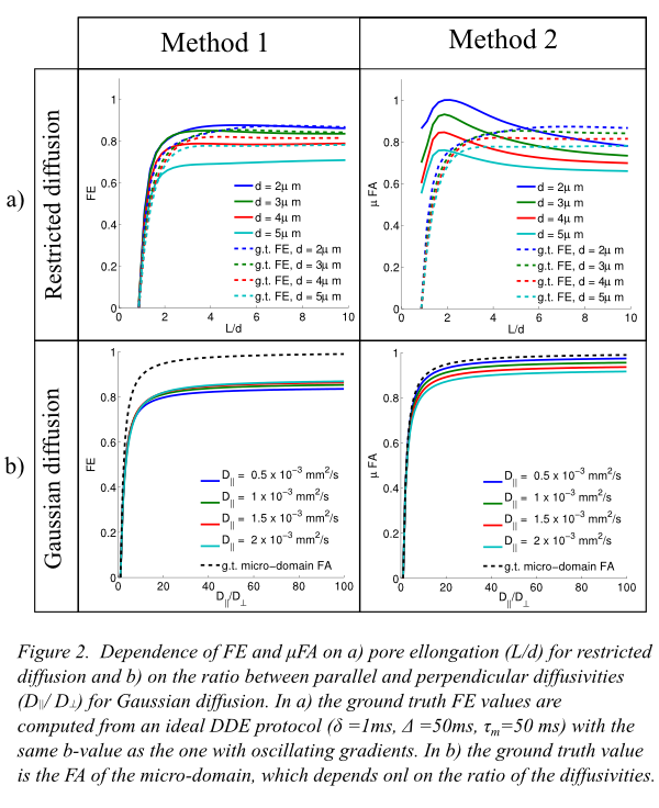
Results: Figure 2 illustrates the dependence of FE and μFA on pore elongation a) and ratio between parallel and perpendicular diffusivities b). For restricted diffusion, FE closely follows the ground truth values, while μFA over estimates them for pores with low eccentricity. Moreover, μFA is not a monotonously increasing function of pore eccentricity. For substrates with Gaussian diffusion, which is the case investigated in [2], μFA is closer to the ground truth FA values then FE. This can be a consequence of using multiple b-values when deriving this metric as opposed to a single b-value for FE.
Discussion: The results show that FE and μFA do not provide exactly the same measure of microscopic anisotropy. For substrates featuring restriction, FE is more accurate, while for substrates featuring Gaussian diffusion μFA is closer to the ground truth.
- [1] S. N. Jespersen, H. Lundell, C. K. Sonderby, and T. B. Dyrby, (2013), Orientationally invariant metrics of apparent compartment eccentricity from double pulsed field gradient diffusion experiments., NMR in Biomedicine, 26:1647-1662
- [2] S. Lasic, F. Szczepankiewicz, S. Eriksson, M. Nilsson, and D. Topgaard, (2014), Microanisotropy imaging: quantification of microscopic diffusion anisotropy and orientational order parameter by diffusion MRI with magic-angle spinning of the q-vector, Frontiers in Physics
