π Echo-Planar Imaging with Concomitant Field Compensation and Efficient k-space Sampling for Porous Media MRI
- University of New Brunswick, Fredericton, Canada
The π Echo Planar Imaging (PEPI) method [1] is a modification of EPI, incorporating a CPMG sequence to utilize RF refocusing instead of gradient reversal. The PEPI method is advantageous for short T2* species where recurrent gradient reversals are not possible. However, it is notoriously difficult to implement due to non-ideal RF pulses and mismatched gradients.
We employ composite broadband refocusing RF pulses and XY phase cycling to compensate for B1 field inhomogeneity and to suppress residual spurious echo signals. The gradient pre-equalization method [2] is employed to ensure high quality gradient waveforms and fast gradient switching. Echo spacings as short as 1.2 ms were achieved for Cartesian line sampling, enabling rapid 3D imaging of short relaxation time species with sub-millimeter resolutions.

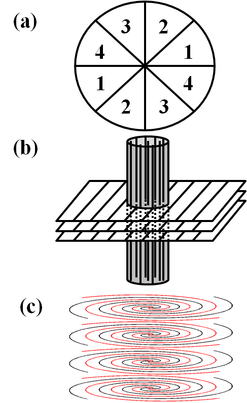
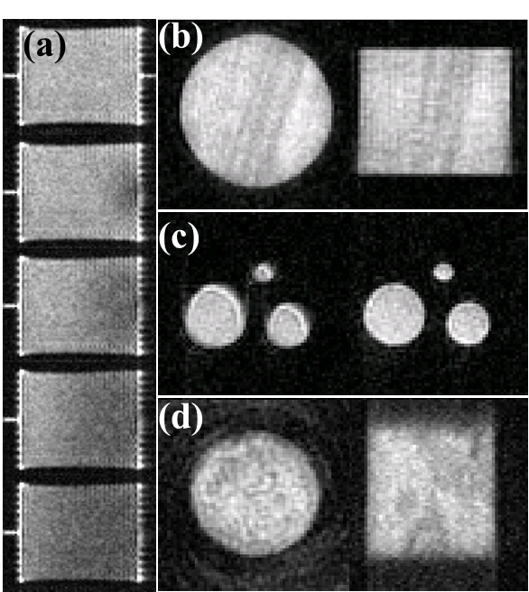
Accelerated PEPI measurements with a variable number of centric interleaves are presented. Fig.1a illustrates the generalized phase encoding scheme with 4 RF excitations. The interleaved scan is suitable for samples with T2* as short as a few hundred microseconds and T2 in the range of tens of milliseconds to hundreds of milliseconds. This scheme provides high temporal resolution in monitoring dynamic systems by simple view sharing. In a continuous acquisition, images at different time points can be obtained by shifting in steps of one or more interleaves. Fig. 2a shows 2D slices of 3D PEPI images monitoring D2O flooding of an oil saturated Bentheimer rock core plug, with 8 centric interleaves acquisition. The temporal resolution was 1.4 min. Every 16th frame is shown. The spatial resolution was 0.75 mm × 0.75 mm × 0.98 mm.

Restricted k-space sampling [4] , as in Fig. 1b, was implemented for cylindrical geometry samples. Good quality images can be obtained with only 15% of the k-space data acquired in 3D imaging. 2D slices of a 3D restricted sampling PEPI image on a Locharbriggs sandstone core plug are shown in Fig. 2b. The spatial resolution was 0.75 mm × 0.75 mm × 0.98 mm. All MR visible structures within the object are well preserved.
Compensation for concomitant magnetic fields by waveform symmetrization [3] was incorporated in the PEPI sequences, which is especially important for applications with high spatial resolution and/or long echo trains. Removal of artifacts due to concomitant magnetic fields is presented in Fig. 2c by comparing the PEPI without and with concomitant field compensation. The shown slices have a spatial resolution of 0.1 mm × 0.1 mm.
For samples with moderate T2*, on the order of several milliseconds, spiral readout is incorporated for efficient sampling, as in Fig. 1c. A spiral-in and spiral-out scheme is employed to acquire the center of k-space at the center of echo signal. The number of spirals is flexible and can be chosen based on sample T2*. Multifrequency reconstructions can be performed if necessary. Fig. 2d shows 2D slices of a 3D image on a Portland sandstone acquired with stack of spirals PEPI. The spatial resolution was 0.8 mm × 0.8 mm × 0.8 mm.
For porous media studies where the sample is limited to the RF probe sensitive region, the broadband 180° RF refocusing quality is usually sufficient through combining composite pulses and phase cycling. Under this condition, the PEPI methods yield superior quality images, compared to the Fast Spin Echo methods. FSE requires high gradient duty cycle and recurrent gradient switchings inducing eddy currents and concomitant magnetic fields. These adverse features are severe on low static field MRI systems (0.2 T) for petroleum reservoir rock core studies, where the concomitant magnetic field will be more than an order of magnitude higher than presented in this work. Rapid PEPI techniques have great potential for monitoring dynamic processes on low field systems.
The π Echo Planar Imaging (PEPI) method [1] is a modification of EPI, incorporating a CPMG sequence to utilize RF refocusing instead of gradient reversal. The PEPI is advantageous for short T2* species where recurrent gradient reversals are not possible.
We employ composite broadband refocusing RF pulses and XY phase cycling to compensate for B1 field inhomogeneity and to suppress residual spurious echo signals. The gradient pre-equalization method [2] is employed for fast gradient switching. Echo spacings as short as 1.2 ms were achieved for Cartesian line sampling, enabling rapid 3D imaging of short relaxation time species with sub-millimeter resolutions. Compensation for concomitant magnetic fields by waveform symmetrization [3] was incorporated in PEPI. Artifacts due to concomitant fields were investigated.

Accelerated PEPI measurements with a variable number of centric interleaves are presented. Fig.1a illustrates the generalized phase encoding scheme with 4 RF excitations. Restricted k-space sampling [4] , as in Fig. 1b, was implemented for specific sample geometries, notably a Locharbriggs sandstone core plug. Good quality images can be obtained with only 15% of the k-space data acquired in 3D imaging. These methods are suitable for samples with T2* as short as a few hundred microseconds and T2 in the range of tens of milliseconds to hundreds of milliseconds.
For samples with moderate T2*, on the order of several milliseconds, spiral readout is incorporated for efficient sampling, as in Fig. 1c. A spiral-in and spiral-out scheme is employed to acquire the center of k-space at the center of the echo signal.
For porous media studies where the sample is limited to the RF probe sensitive region, the broadband 180° RF refocusing quality is usually sufficient through combining composite pulses and phase cycling. Under this condition, the PEPI methods yield superior quality images, compared to the Fast Spin Echo methods. FSE requires a higher gradient duty cycle and recurrent gradient switchings inducing eddy currents and concomitant magnetic fields. These adverse features are severe on low static field MRI systems (0.2 T) for petroleum reservoir rock core studies, where the concomitant magnetic field is more than an order of magnitude higher than presented in this work. Rapid PEPI techniques have great potential for monitoring dynamic processes on low field systems.
- [1] D. N. Guilfoyle, P. Mansfield, (1992), Fluid flow measurement in porous media by echo-planar imaging., Journal of Magnetic Resonance, 97, 342-358
- [2] F. G. Goora, B. G. Colpitts, B. J. Balcom, (2014), Arbitrary magnetic field gradient waveform correction using an impulse response based pre-equalization technique., Journal of Magnetic Resonance, 238, 70-76
- [3] X. J. Zhou, S. G. Tan, M. A. Bernstein, (1998), Artifacts induced by concomitant magnetic field in fast spin-echo imaging., Magnetic Resonance in Medicine, 40, 582-591
- [4] D. Xiao, B. J. Balcom, (2013), Restricted k-space sampling in pure phase encode MRI of rock core plugs., Journal of Magnetic Resonance, 231, 126-132
