Magnetic Resonance Imaging of Idealised Wetland samples produced using Additive Manufacturing
- 1. Nottingham Trent University, Physics and Mathematics, Nottingham, United Kingdom
- 2. BASF SE Materials Physics and Analytics, Ludwigshafen, Germany
The disruption to MR images caused by susceptibility artefacts is well known within the porous media community and makes it very challenging to undertake imaging of samples containing ferromagnetic material. Although not necessarily a key constituent of many gravels, some particles within the mixture will contain large quantities of iron, making it difficult if not impossible to guarantee a good quality image. Packs of glass beads have long been used as a model for such systems and although representative, the results may not reveal interesting features which the random shapes of gravel particles produce. Furthermore, loose glass beads are prone to movement and certainly in changes of sample orientation whilst fused glass beads lead to less well defined geometry. In this work, we present a new technique by which ABS replicas of gravel packs can be produced and MR images collected without the difficulties encountered with 'dirty' gravel. For comparison, these results are compared to those from ABS bead packs of different sizes.
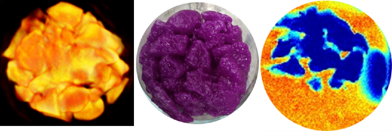
For the bead packs, perfect hexagonal packing is achieved by allowing the beads to pass into the outer shell. For the replica gravel samples, gravel packs are first scanned with X-ray CT which is used to produce spatial information from which the gravel packs are made. MRI is conducted in each of these replica samples and in the original gravel pack. The modelled samples are produced on a CTC Replicator clone from ABS. The printed structure is treated with acetone to make it waterproof. CT imaging is used on the final prints as well as proton density images to allow comparison with the original structure.

It is found that the gravel sample is too magnetic to be imaged. The replica sample provides excellent image quality. Of particular interest is the behaviour of wastewater passing through these systems as models for constructed wetlands. To explore this, each of the idealised systems is imaged before and during flow with wastewater. Over a period of several weeks, biofilms establish MR relaxation images reveal the distribution of water, particulate matter and biofilms in the samples.
For the first time we have printed an ABS replica of a gravel sample which was scanned using X-Ray CT. We have shown that by doing this, the build-up of biofilms and particulates can be assessed in a sample which has the same structure as a genuine gravel sample but without the issues associated with susceptibility artefacts. Validation of a possible impact of different surface chemistry on the spatial pattern of biofilm growth is under way.
