Diffusion tensor imaging of a chemically fixed CS23 human embryo at 9.4 T
- 1. University of Tsukuba, Institute of Applied Physics, Tsukuba, Japan
- 2. Kyoto University, Graduate School of Human Health Sciences, Kyoto, Japan
Diffusion tensor imaging (DTI) is now widely used for visualization of neuronal structure of human fetal brains. However, as far as we know, DTI measurements have been limited more than 13 weeks after conception. In this study, we performed DTI of a Carnegie Stage (CS) 23 (8 weeks after conception) chemically-fixed embryo using a 9.4 T MRI system.
A CS 23 chemically-fixed human embryo was selected from the Human Embryo Collection preserved in Kyoto University. The embryo was stored in an NMR sample tube (15 mm OD and 13.5 mm ID) filled with formalin solution. The test tube was inserted into a birdcage RF coil (diameter = 18 mm, length = 28 mm) tuned for 400 MHz. MR images of the specimen were acquired using a home-built digital MRI system using a 9.4T/54mm vertical bore superconducting magnet. Standard six-axis diffusion-weighted imaging sequences (TR=1200ms, TE=32ms, 2NEX, δ =6ms, Δ =16ms, b=790 s/mm2, image matrix = 128x128x256, voxel size=(120 μ m)3) were used.
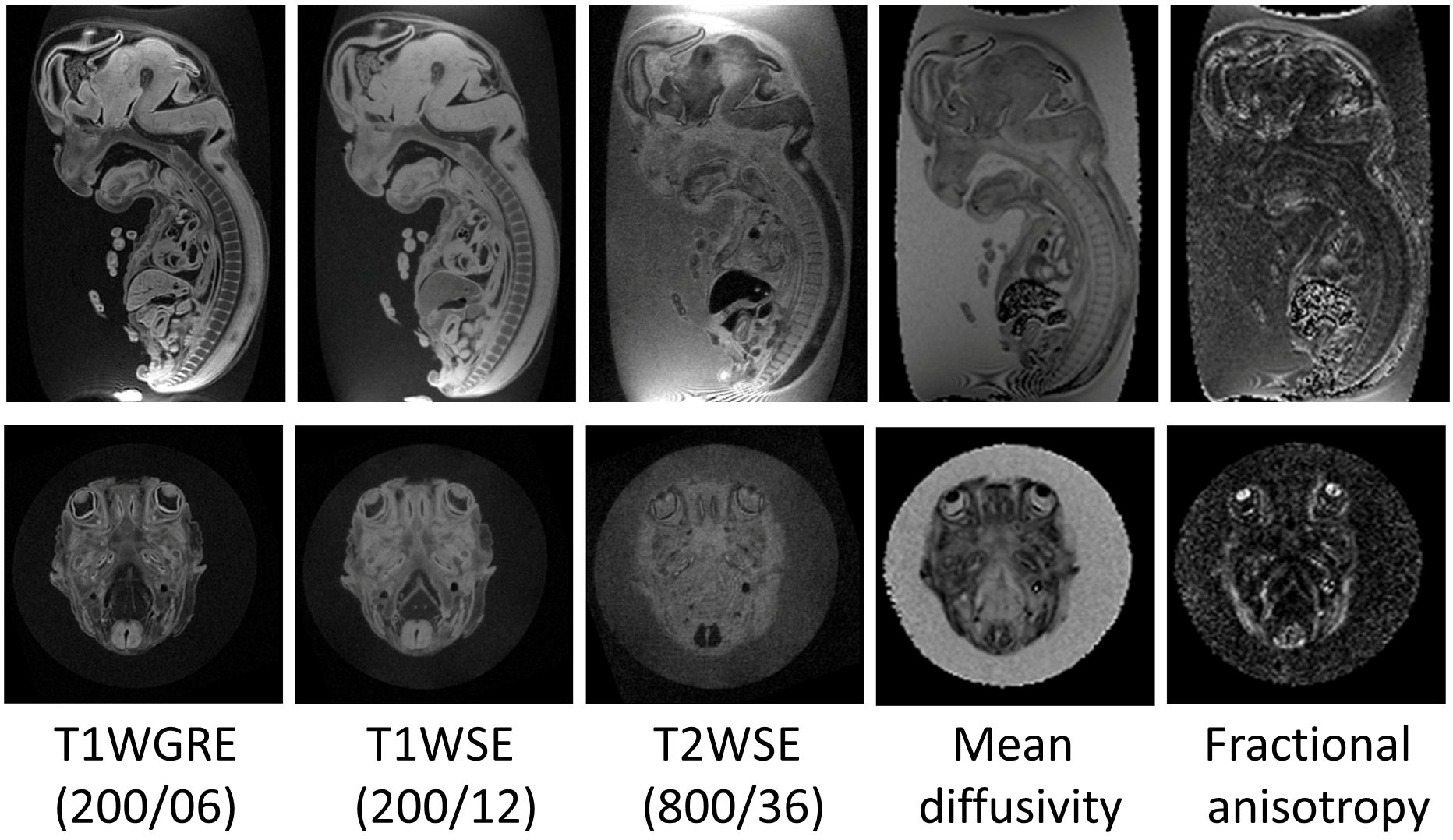
Figure 1 shows the mid-sagittal cross-section and the transverse cross-section at the eye-level selected from 3D image datasets acquired with a T1-weighted gradient-echo sequence (TR/TE=200ms/6ms), T1-weighted spin-echo sequence (TR/TE=200ms/12ms), and T2-weighted spin echo sequence (TR/TE=800ms/36ms), and 3D datasets of mean diffusivity (MD) and fractional anisotropy (FA) calculated from the diffusion weighted images. These images clearly show that the lens have shorter T1, small MD, and large FA, which suggests the tissue of the lens is anisotropic. In conclusion, DTI of a chemically fixed human embryo presents new information for its microstructure.

