High resolution fMRI: a technique to investigate single cell behavior
- Centre CEA de Saclay (Essonne), Gif sur Yvette, France
Despite the tremendous advancements made in the field of magnetic resonance imaging (MRI) in general, and of functional MRI (fMRI) in particular, most functional neuroimaging investigations are limited to averaging the signal from clusters of hundreds of neurons. In the traditionally used blood-oxygen-level-dependent fMRI (BOLD-fMRI) approach the spatial resolution is limited by the maximum affordable acquisition time and by the mechanism governing the BOLD effect which detects neuronal activity indirectly through blood flow changes.
In the last decade, a new fMRI technique, manganese-enhanced MRI (MEMRI), has been successfully proven on various vertebrate animal models. MEMRI uses manganese, an MR contrast agent, to label active neurons. The amount of manganese that accumulates intracellularly is directly linked to neuronal activity, because the Mn2+ ion enters the neurons through calcium and nonspecific cationic channels. Unlike BOLD-fMRI, MEMRI measures changes in calcium-dependent activity independently of hemodynamic effects, and allows brain imaging after the active behavior, permitting therefore higher spatial resolutions.
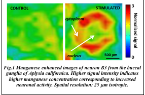
Here we demonstrate the feasibility of performing functional MEMRI studies with single-cell resolution. At ultrahigh magnetic field, manganese-enhanced magnetic resonance microscopy allows the identification of most motor neurons in the buccal network of Aplysia at low, nontoxic Mn2+ concentrations. We establish that Mn2+ accumulates intracellularly on injection into the living Aplysia and that its concentration increases when the animals are presented with a sensory stimulus (Fig.1). We also show that we can distinguish between neuronal activities elicited by different types of food-related stimuli (arousing and rewarding). This method opens up a new avenue into probing the functional organization and plasticity of neuronal networks involved in goal-directed behaviors with single-cell resolution.

